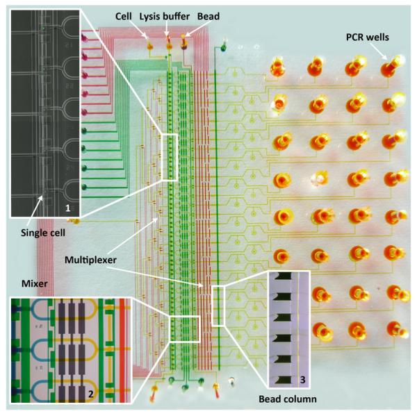
Fig. 1.

A microphotograph of a multilayer microfluidic chip designed for single-cell analysis. All the microfluidic channels were filled with different food dyes, indicating flow channels (yellow), control valves (green), and multiplexer control channels (red), respectively. Inset 1 is a composite image indicating five single-cells individually captured in the cell loading units. Inset 2 is the cell lysis module. Cell lysis is performed by opening the portion valve and pumping to mix lysis buffer (yellow) with the captured cell (blue). Two sets of multiplexers make each cell manipulation unit and bead column individually addressable for high throughput analysis. Inset 3 shows six stacked oligo-dT bead columns next to the sieve valve. The cell lysate was pushed through oligo-dT bead columns for mRNA capture and cDNA synthesis. Finally, the beads with the synthesized cDNA were collected in the PCR wells in the chip substrate for PCR analysis.
