Fig_1.

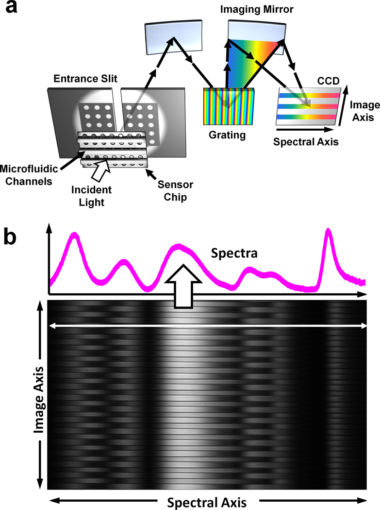
Spectral imaging setup. (a) An imaging spectrometer is used to acquire multiple transmission spectra from a single nanohole array chip with 50 parallel microfluidic channels. An image of the transmitted light is focused onto the entrance slit, with perpendicular microfluidic channels. This light is dispersed along one dimension, forming a spectral image on a CCD chip. The vertical, or imaging, axis corresponds to position along the entrance slit whereas the horizontal axis corresponds to the spectral content of the light entering the slit. (b) Sample data recorded on the CCD. The microfluidic channels are clearly seen spaced vertically. A single horizontal cross section line shows the transmission spectrum from a single channel.
