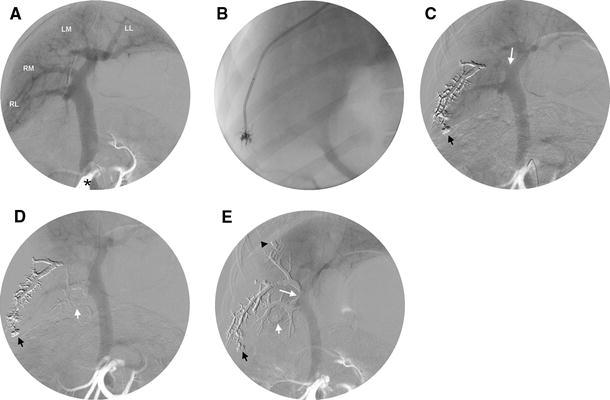
Fig. 1.

A Preembolization portogram showing the portal supply to the right lateral lobe (RL), the right medial lobe (RM), the left medial lobe (LM), and the left lateral lobe (LL), and the position of the arterial catheter in the cranial mesenteric artery (*). B Injection of Onyx from a wedged catheter position in the hepatic vein. C Onyx cast is visible at portography after first injection (injection position marked by the black short arrow). This injection was unsuccessful, because the segmental portal vein was not embolized up to its origin (white long arrow). D Onyx cast during the second injection (injection position marked by the white short arrow) successfully embolizing the RL portal vein. E Final portography shows a successfully embolized right hemiliver after the RM portal vein had been embolized by the third injection (injection position marked by the black arrowhead). Note that the Onyx cast from the second injection extends into the portal trunk (white long arrow). The diameter of the portal vein has markedly decreased compared with the preembolization portogram (A)
