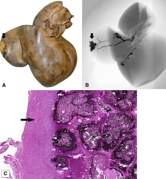
Fig. 3.

A Macroscopic view of a capsular lesion (arrow), consistent with B a superficial injection site (arrow) under fluoroscopy. C Microscopic view (H&E, original magnification ×40) of the same region shows capsular fibrosis (arrow) around the injection site
