
Figure 1. Identification of rat aortic body and carotid body cell types in situ and/or in dissociated cell culture.
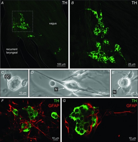
A, confocal section of a whole-mount containing the vagus and recurrent laryngeal nerve bifurcation reveals aortic body type I cell clusters, immunostained for tyrosine hydroxylase (TH); inset in A shown at higher magnification in B. C–E, phase contrast micrographs of dissociated AB cells after 24 h in culture. An isolated AB type I or glomus cell (GC) cluster is shown in C; the soma of local neurons (N) with and without visible processes are shown in D and E, respectively. Parallel cultures of dissociated AB cells (F) and CB cells (G) from the same animals, showing type I clusters immunopositive for TH (green), and glia-like cells, immunopositive for glial-fibrillary acidic protein (GFAP, red). Note GFAP-positive cells are more abundant in AB cultures (F), relative to CB cultures in which GFAP is known to stain predominantly glia-like, type II cells (G).