
Table 1.
MR Imaging R2* Values Compared with Histologically Determined Iron Grades
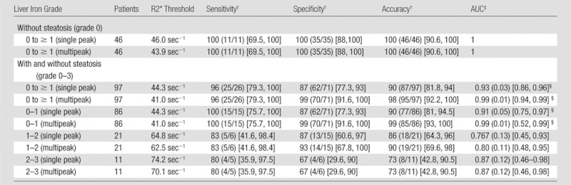
Note.—After exclusion of all patients with steatosis grades of 1 or more (first rows), there were no significant differences of AUC, and excellent sensitivity, specificity, accuracy were observed when single-peak and multipeak reconstruction were used for histologically determined iron grade 0 to ≥ 1. In comparison, there was a significant difference in AUC for single-peak reconstruction if samples with fat were included (P = .005). Iron quantification with R2* mapping is confounded by the presence of fat when single-peak reconstruction is used. Threshold values for different histologic iron grades are defined in the third column.
Data are percentages, with numerator and denominator in parentheses and 95% confidence interval in brackets.
Data in parentheses are standard error, with 95% confidence interval in brackets.
Indicates a statistically significant difference, P <.05.