
Figure 1.
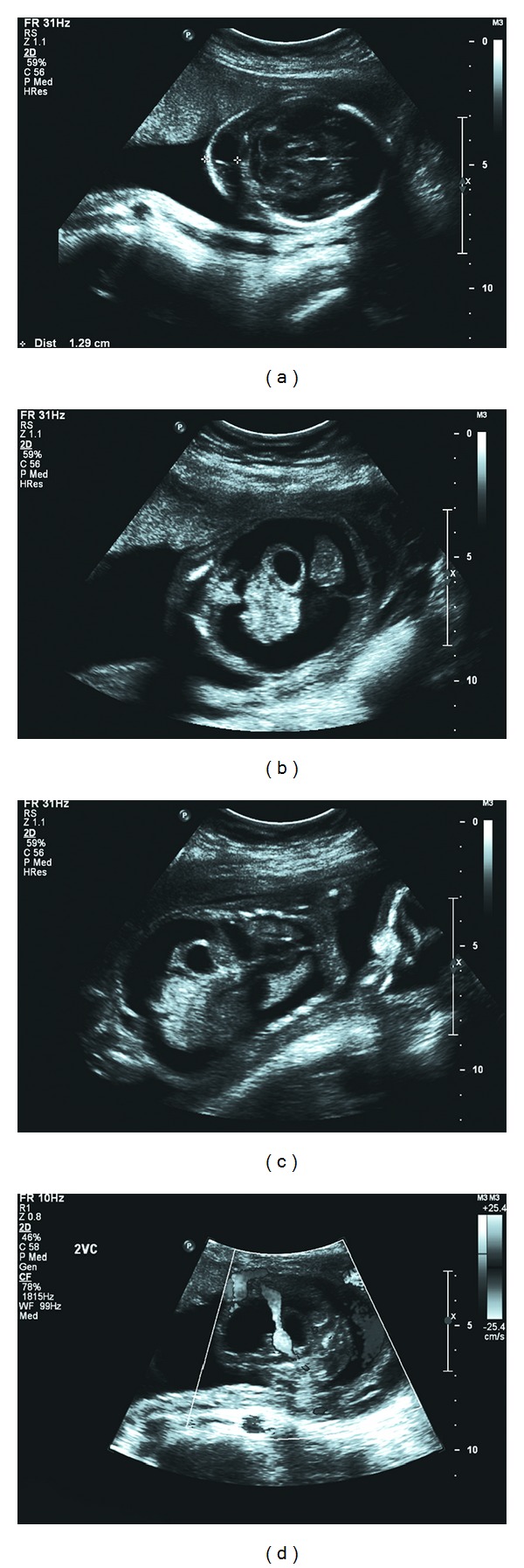
Ultrasound images of (a) septated cystic hygroma, (b) ascites with free floating stomach and small intestine, (c) ascites and pleural effusions (coronal view), and (d) single umbilical artery.
Official websites use .gov
A
.gov website belongs to an official
government organization in the United States.
Secure .gov websites use HTTPS
A lock (
) or https:// means you've safely
connected to the .gov website. Share sensitive
information only on official, secure websites.
Ultrasound images of (a) septated cystic hygroma, (b) ascites with free floating stomach and small intestine, (c) ascites and pleural effusions (coronal view), and (d) single umbilical artery.