Figure 2.
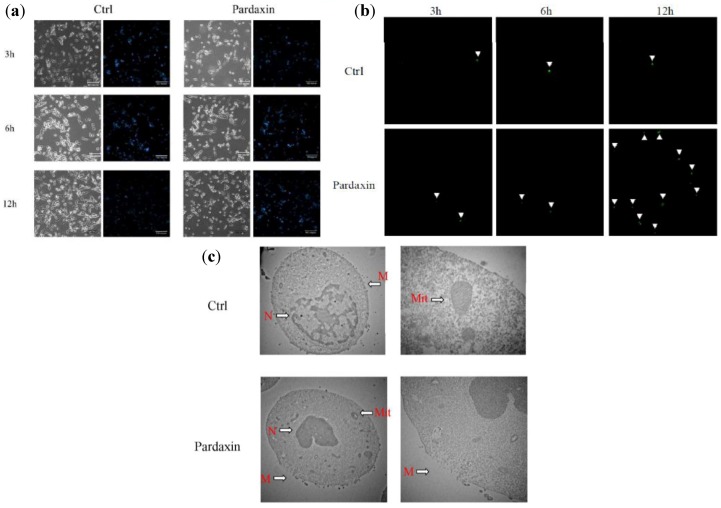
Morphological changes in MN-11 cells after exposure to different concentrations (0 and 13 µg/mL) of pardaxin for 3, 6, and 12 h. (a) Detection of typical features of apoptotic nuclear condensation by Hoechst 33258 staining (magnification 200×). (b) Caspase-3/7 activity was measured after 3, 6, and 12 h of treatment, and green color was detected under a fluorescence microscope. The green color is indicated by white arrowheads. (c) Effects of pardaxin on membranes of MN-11 tumor cells examined by transmission electron microscopy. Untreated cells (Ctrl) showed a normal surface, while cells treated with pardaxin (13 µg/mL) for 24 h revealed disrupted cell membranes. N, nuclear; M, cell membrane; Mit, mitochondria.

