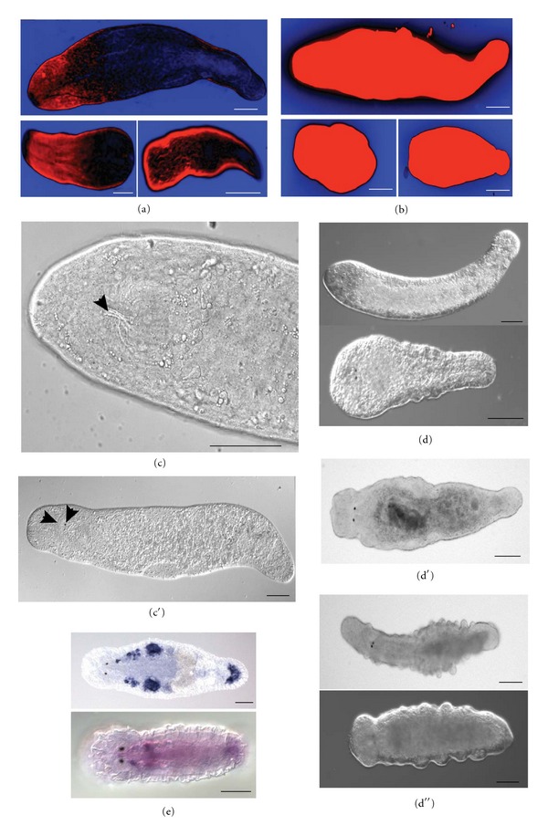
Figure 3.

Bioelectric signaling and stem cells in M. lignano. (a-b) DiBAC4(3) staining of intact worm (top), anterior (left bottom) and posterior (right bottom) fragments. (a) control worm, (b) worm treated with 1 μM IVM. Blue is more polarized than black, black is more polarized than red. (c-c′) Regeneration of head-specific structures after 1 μM IVM treatment. Arrowheads in (c) indicate regenerated pharynx, in (c′) regenerated eye and half of the brain. (d-d′′) intact worms exposed to high doses of IVM (2 μM in d and 4 μM in d′) and PZQ (150 μM in d′′). (d) head regression; (d′) square head; (d′′) bulges and outgrowth. (e) In situ hybridization results in adult (top) and juvenile (bottom) animals with the probe against RNA815_5834 transcript from ML110815 transcriptome assembly (voltage-gated sodium channel). In juvenile worm this gene is expressed almost ubiquitously, and in adults expression is only detected in gonads and (likely) in somatic stem cells. Strong signal in the adhesive glands in the tail is likely a common artifact.
