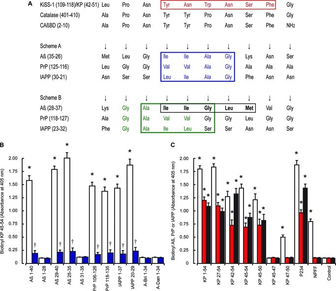
Figure 7.
Binding of KP to Aβ, PrP and IAPP peptides. Alignment of the human metastasis-suppressor KiSS-1 preproprotein sequence (NP_002247.3) with the human catalase sequence (NP_001743.1) is shown in A. The red box highlights the region of KP that prevents Aβ, PrP, and IAPP toxicity; blue and green boxes highlights the Gly-Ala-Ile-Ile region that binds catalase in schemes A and B respectively; the black box highlights Aβ 31–35, which inhibits Aβ 1–42 binding to catalase. Immunoplates were coated with Aβ 1–40, Aβ 1–28, Aβ 29–40, Aβ 25–35, PrP 106–126, PrP 118–135, IAPP 1–37, IAPP 20–29, A-Bri 1–34, or A-Dan 1–34 fibrils. Coated plates were incubated with either biotinylated KP 45–54 (B) alone (open columns) or in the presence of unlabeled KP 45–54 (closed blue columns) and bound material determined by EIA. Plates coated with KP 1–54, KP 27–54, KP 42–54, KP 45–54, KP 45–50, KP 45–47, KP 47–50, or NPFF (C) were incubated with biotinylated Aβ 1–42 (open columns), biotinylated PrP 106–126 (red columns), or biotinylated IAPP 1–37 (black columns) and bound material determined by EIA. All results are expressed as the mean ± SEM (n = 8). (* = P < 0.05 vs control (buffer alone); † = P < 0.05 vs biotinylated KP 45–54; one-way ANOVA.)

