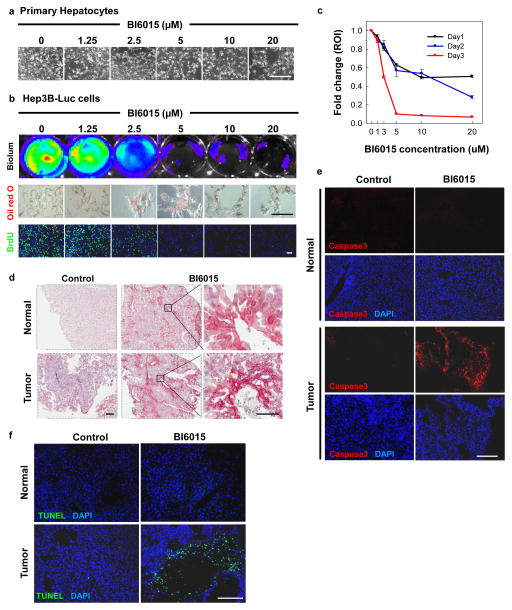
Figure 6. HNF4α antagonists are cytotoxic to hepatocellular carcinoma.
(a) Toxicity was not observed in primary hepatocytes treated with BI6015 in vitro. (b) BI6015 was potently toxic to Hep3B-Luc cells, as measured by bioluminescence. Fat accumulation, measured by Oil Red O staining (red), was enhanced in Hep3B-Luc cells treated with BI6015, while cell division, measured by BrdU uptake (green), was blocked in a dose-dependent manner. (c) Bioluminescence of Hep3B-Luc treated with BI6015 was quantified at 24, 48 and 72 hours after treatment. Decreased bioluminescence correlated directly with decreased cell counts. (d) Hep3B-Luc cells were injected into the liver parenchyma of nude mice, hereafter referred to as HCC mice. Animals were injected IP with 30 mg/kg of BI6015 daily or every other day, as tolerated. Treatment with BI6015 resulted in marked induction of Oil Red O staining (red) in both the normal liver and tumor region, as compared with DMSO. (e and f) Apotosis was evaluated by immunostaining for cleaved caspase-3 (red) and a TUNEL assay (green). High levels of cleaved caspase-3 expression were found in the tumor regions, but not in the normal liver of BI6015-treated HCC mice as compared with the DMSO control. A similar phenomenon was observed with TUNEL positive cells, which were only found in tumor regions of BI6015-treated HCC mice, predominantly in regions surrounding the necrotic core of the tumor. Scale bars, a,d, e,f=200 μm (inset for d=50 μm), b=100 μm.

