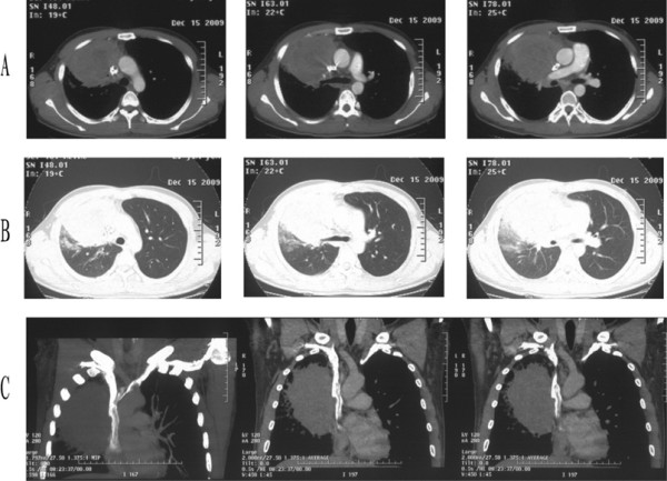
Figure 1.

Enhanced chest CT scan before operation. (A) In the mediastinal window, the CT scan revealed the tumor encroaching on the superior vena cava (right panel), surrounding the right upper lobe bronchus (middle panel), and invading the right pulmonary artery (left panel). (B) In the lung window. (C) In the mediastinal window, coronal.
