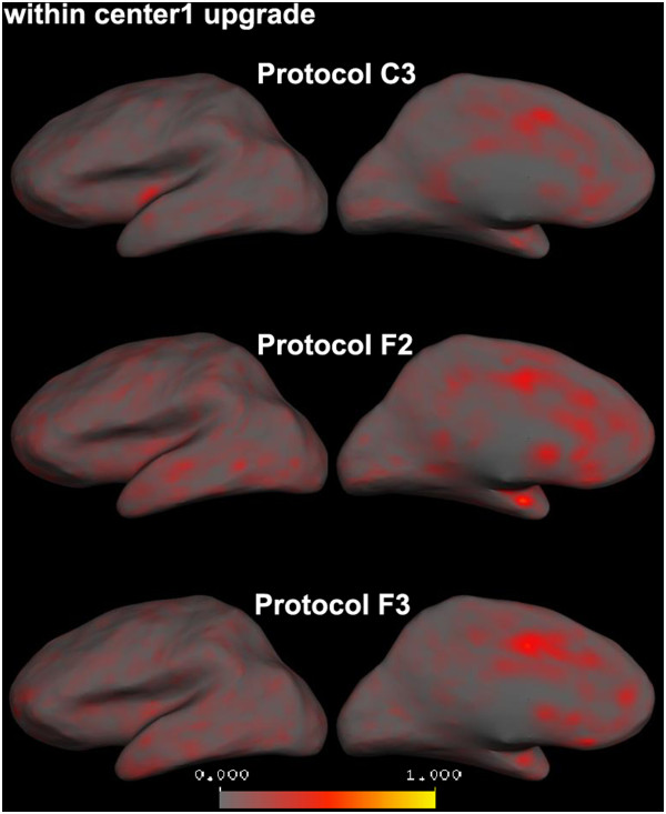
Figure 7.

Average absolute cortical thickness differences between before and after scanner upgrade images in Center1 calculated for each vertex on the left cortical surface (right hemisphere is similar). All the protocols show low average absolute cortical thickness differences and therefore high reproducibility.
