Figure 1.
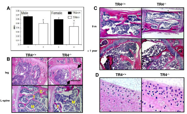
Skeletal abnormalities in TR4−/−mice.A: Reduced bone mineral density in TR4−/− mice, in both genders. DEXA scan of 6 pairs and 5 pairs of 6 months old TR4−/− male and female mice, respectively. The average bone density was compared. B: Bone histology. Reduced bone volume in TR4−/− long bone (upper panel) and spinal column (L-spine, lower panel). HE staining is from 6 month old TR4+/+ and TR4−/− mice legs and lumbar-spines. M: bone marrow; T: trabeculae; C: cortex. Accumulation of adipocytes found in TR4−/− mice. Arrows indicate that there is more fat accumulation in TR4−/− mice bone marrow. C: Continuing reduced bone volume at 9 months and over one year old TR4−/− mice. D: Osteoarthritis in TR4−/− mice. 6 months old TR4−/− knee joint shows signs of osteoarthritis.

