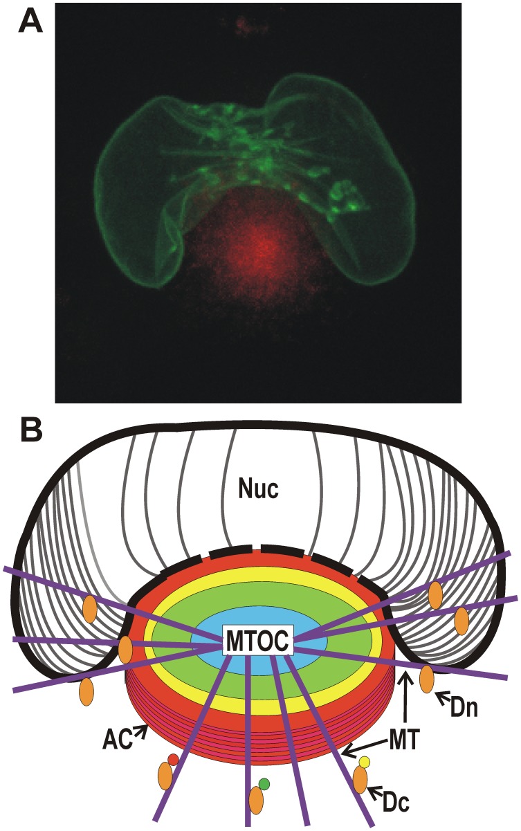
Figure 1. Microscopic and diagrammatic representations of the assembly compartment and nucleus in an HCMV-infected cell.
(A) Maximum projection live cell micrograph showing the AC (identified by Dsred-tagged pp28, red) with the enlarged, kidney-shaped nucleus (identified by GFP-tagged lamin A) wrapped around the AC. (B) A poor artist's diagrammatic representation of the AC located next to the enlarged, kidney-shaped nucleus. The AC is formed on a microtubule organizing center (MTOC) with microtubules (MT) radiating from it. The nested cylindrical makeup of the AC is indicated by colored circles; each cylindrical region is proposed to be made up of vesicles derived from specific secretory organelles. Virion tegument and structural proteins reside with these vesicles and are applied to nucleocapsids as they egress from the nucleus and traverse toward the center of the AC. The point of contact between the AC and the nucleus may be a continuum, suggested by the dashed line representing the nuclear membrane at this junction. This may allow nucleocapsids open access from the nucleus to the AC late in infection. The AC and the kidney-shaped nucleus are formed by virally commandeered functions of dynein. Dynein (Dn) is shown pulling the nucleus around the AC by a mechanism similar to mitotic nuclear envelope breakdown. Dynein loaded with cargo (Dc) is shown to represent the formation of the AC.

