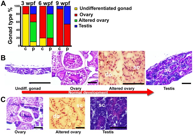
Figure 7. Gonad development.
A: Gonad type percentage of 10 zebrafish from control (c) and probiotic (p) groups at 3, 6 and 9 weeks post fertilization – wpf. Assay was run in triplicate. B: Normal gonad development; progression from undifferentiated gonad, to ovary, altered ovary and testis. C: representative pictures of ovary, altered ovary and testis prematurely observed in probiotic treated fish at 3 weeks post fertilization. Legend: go, gonocyte; do, degenerating oocytes; sc, spermatocyst; I, stage I oocyte; II, stage II oocyte; scale bars 50 µm.

