
Figure 1. FRET-based conformational sensor for the m1 AChR.
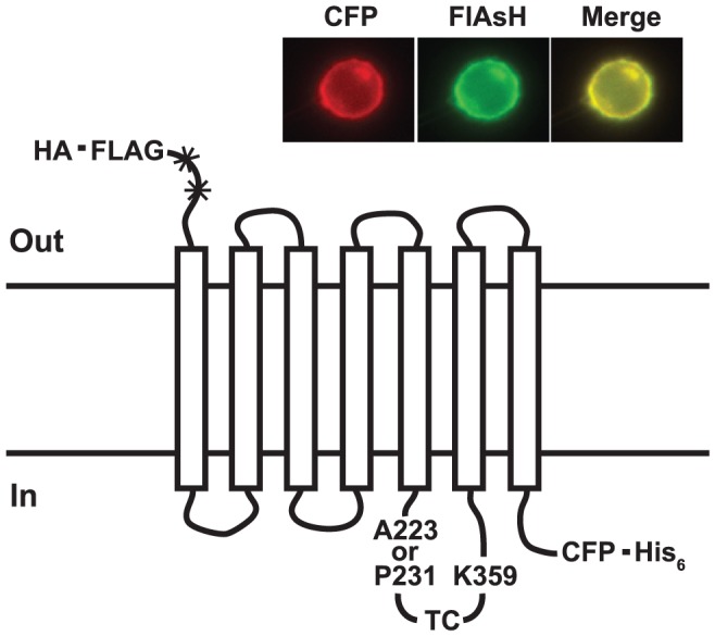
A CFP FRET donor is placed at the C terminus before a His6 purification tag. A TC motif (CCPGCC), the labeling site for the FlAsH FRET acceptor, is inserted in the i3 loop between the sites shown. Details are given in the text. Glycosylation sites (*) are destroyed and a HA signal sequence and FLAG epitope are placed at the N terminus. Inset: Subcellular location of one of the prototype sensors in HeLa cells viewed with a filter set for either CFP or FlAsH.