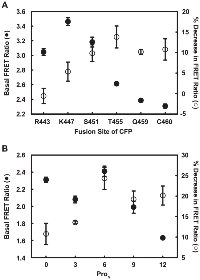
Figure 2. Optimization of the C terminal CFP fusion site.

The position of the CFP FRET donor was varied either (A) by truncation of C-terminal residues before the attachment site or (B) by introduction of oligo-proline spacers (Pron) after C460, the native C terminus. Membranes prepared from FlAsH-labeled HeLa cells that expressed each variant were used to measure FRET. Basal FRET ratio (close circles) and percent decrease in FRET ratio in response to 1 mM Cch (open circles) were calculated as described in Materials and Methods. Data are averages and standard deviations from triplicate measurements. Similar results were repeated with membranes prepared from another set of labeled cells.
