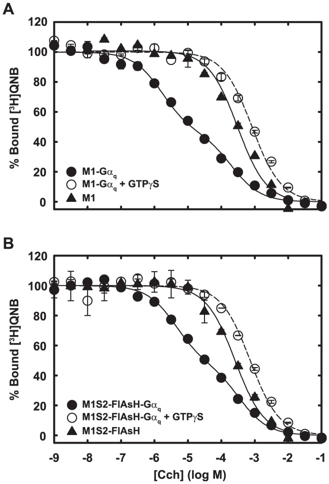
Figure 7. Agonist binding to M1S2-FlAsH and wild-type m1 receptor reconstituted in phospholipid vesicles.

Cch binding to wild-type m1 receptor (A) and in vitro labeled M1S2-FlAsH (B) after their reconstitution into phospholipid vesicles was measured by competition with 2 nM [3H]QNB. Receptors were reconstituted into phospholipid vesicles with (circles) or without (triangle) Gαqβ1γ2. When indicated, 50 µM GTPγS was added in the binding reaction (open circle). Data are averages of duplicate measurements and are expressed as percent of maximum bound [3H]QNB. Error bars indicate the range. Binding in the presence of Gαq (no GTPγS ) was fitted with a two-site binding equation. Binding in the absence of Gαq (filled triangle) or in the presence of added GTPγS (open circle) was fitted with a one-site binding equation.
