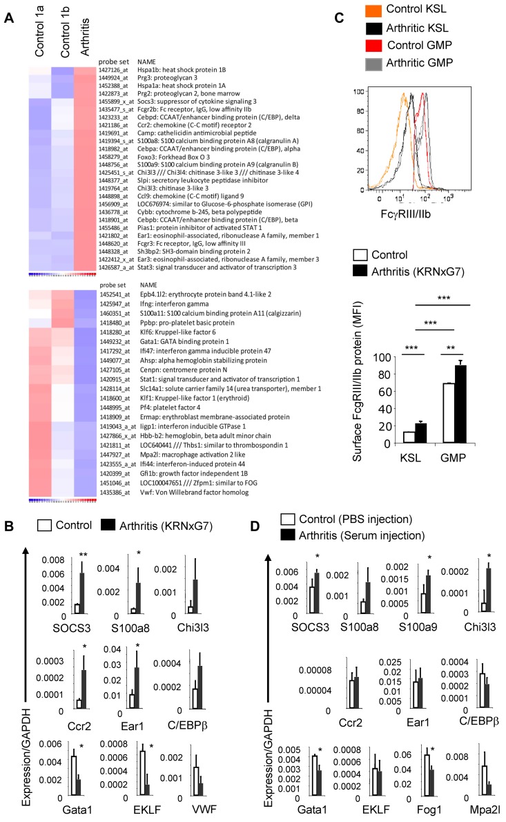
Figure 3.
Arthritic KSL cells show molecular evidence of myeloid priming. (A) Microarray-based heat map depicting selected genes up-regulated (top) or down-regulated (bottom) in arthritic (KRNxG7) KSL cells relative to both B6xG7 (control 1a) and KRN (control 1b) KSL populations. KSL cells from 7-9 mice per strain was used. (B) Quantitative real-time PCR validation of selected differentially regulated genes. Three independent KSL pools each from KRNxG7 arthritic and B6xG7 control mice were used. KSL cells were also independent from pools used for microarray. (C) Top panel, FcγRIII/IIb protein expression on KSL cells and GMP determined by FACS. Histograms from 2 arthritic mice (KRNxG7) and 2 control mice (KRN) are shown. Bottom panel, Median fluorescence intensity (MFI) of anti-FcγRIII/IIb–PE signal on gated KSL cells and GMP as a surrogate quantification of FcγRIII/IIb surface protein level. Representative of at least 2 experiments with at least 3 arthritic and control mice per experimental set up. (D) Quantitative real-time PCR of selected genes in KSL cells from B6 mice that received PBS (control) or serum (developed arthritis). *P < .05, **P < .01, ***P < .001 (see also supplemental Figure 4).

