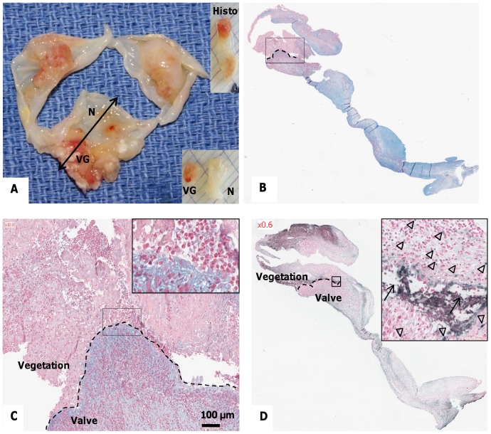
Figure 1. Characterization of the excised valve(s) containing infected vegetations.
(A) Macroscopic view of human endocarditic valves, sectioned for histological analysis (“Histo”) as indicated by the double-headed arrow. Vegetations (VG) and adjacent macroscopically normal (N) parts of the valve were then incubated in culture medium for preparation of “conditioned medium”. (B) Alcian blue staining with nuclear fast red counterstaining of a vegetation and underlying valvular tissue. Blue areas correspond to mucoid degeneration interfacing with infected thrombus (rectangle, magnified in C). Red staining shows inflammatory cell nuclei as well as the septic thrombus. (C) Higher magnifications of the interface between the vegetation and the valvular tissue (Alcian blue staining). Inset shows an accumulation of cells with polylobed nuclei. (D) Detection of fragmented DNA by TUNEL showing apoptotic cells (arrowheads) and extracellular fragmented DNA (arrows). Nuclei were counterstained with nuclear fast red.

