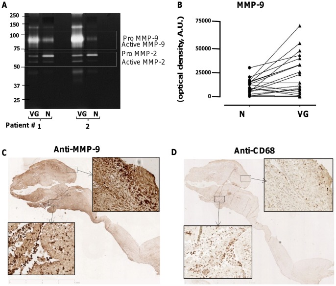
Figure 5. Identification and activity of MMPs and plasmin in human endocarditic valves.
(A) Detection of the different forms of MMP-9 and MMP-2 in conditioned media of VG and N, in representative zymogram from 2 patients. Gelatinolytic bands corresponding to proMMP-9, MMP-9, proMMP-2 and MMP-2 were quantified and expressed in arbitrary optical density units. (B) Diagram illustrating the variation of MMP-9 activity between N and VG for 19 pairs of conditioned media (P<0.01). (C,D) Immunostaining of MMP-9 and macrophages respectively (x1.2) in human endocarditic valves. (C) Strong immunostaining of MMP-9 in the VG and adjacent valve tissue. Insets: higher magnification (x20) of the interface VG/valve (bottom left) and within the valvular tissue (top right). (D) Presence of numerous macrophages at the interface VG/valve where only few of them could be observed in the upper area. Insets: higher magnification (x20) of the interface VG/valve (bottom left) and within the valvular tissue (top right).

