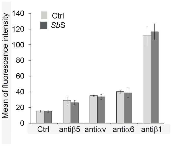
Figure 5. Impact of SbS on integrin expression.
Sub-confluent HCT-8/E11 cells were incubated for 5 h with (dark bars) or without (light bars) SbS, then harvested and resuspended in the presence of anti-integrin subunit mAbs. After incubation with the appropriate secondary Alexa 488-conjugated Ab, cell-bound fluorescence was quantified using a Becton-Dickinson FACScan flow cytometer. Non-specific labeling was determined by incubating cells with the secondary Alexa 488-conjugated Ab alone (Ctrl). Data represent the mean+SD of 6 separate experiments.

