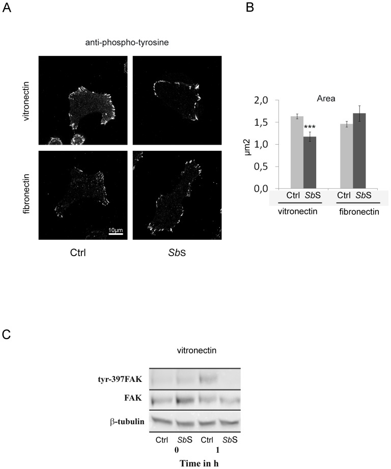
Figure 8. SbS alters the functionality of adherence structures.
(A) HCT-8/E11 cells were plated on either fibronectin or vitronectin for 2 h and then fixed. Tyrosine phosphorylated proteins were stained using a PY20 mAb. (B) Focal adhesion labeling was quantified by measuring the area of each focal adhesion for all cells. Data represent the mean+SD of 3 separate experiments. (C) HCT-8/E11 cells were allowed to adhere on vitronectin after SbS pretreatment. The phosphorylation of tyrosine residues in FAK (Tyr-397FAK) was determined after cell lysis at the indicated times of cell adhesion. Samples were analyzed by western blot analysis. Equal amounts of protein were analyzed and loading amounts were verified by probing the blot with anti-FAK (FAK) or anti- β-tubulin (β-tubulin) Abs. *** P<0.001.

