
Figure 1. V-ATPase localization in the mouse olfactory epithelium.
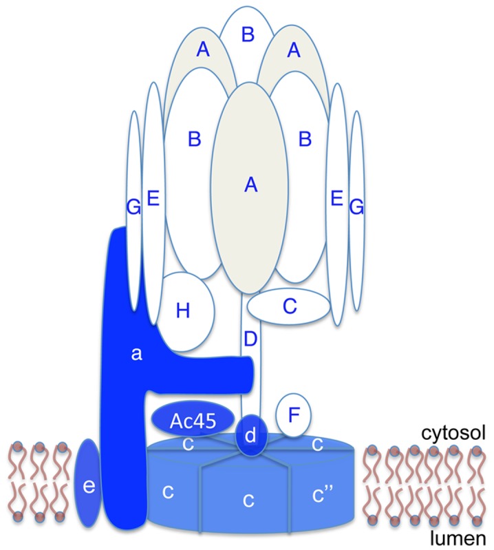
(A) A schematic diagram showing the subunit composition of the V-ATPase. The cytosolic V1 domain is composed of subunits A through H (shown in white or light gray, marked with blue letters). The transmembrane V0 domain is composed of subunits a, c, c” (or b), d, e, and Ac45 (shown in blue, marked with white letters). Some of the subunit interactions are putative. (B) Section from a 3-D image reconstruction showing that B1 V-ATPase (red) localizes to the microvilli of olfactory sustentacular cells in a 2-week old female mouse pup. Apical cilia of olfactory sensory neurons are immunostained for CNGA2 (green). DAPI (blue) stains cell nuclei. Bar = 30 µm.