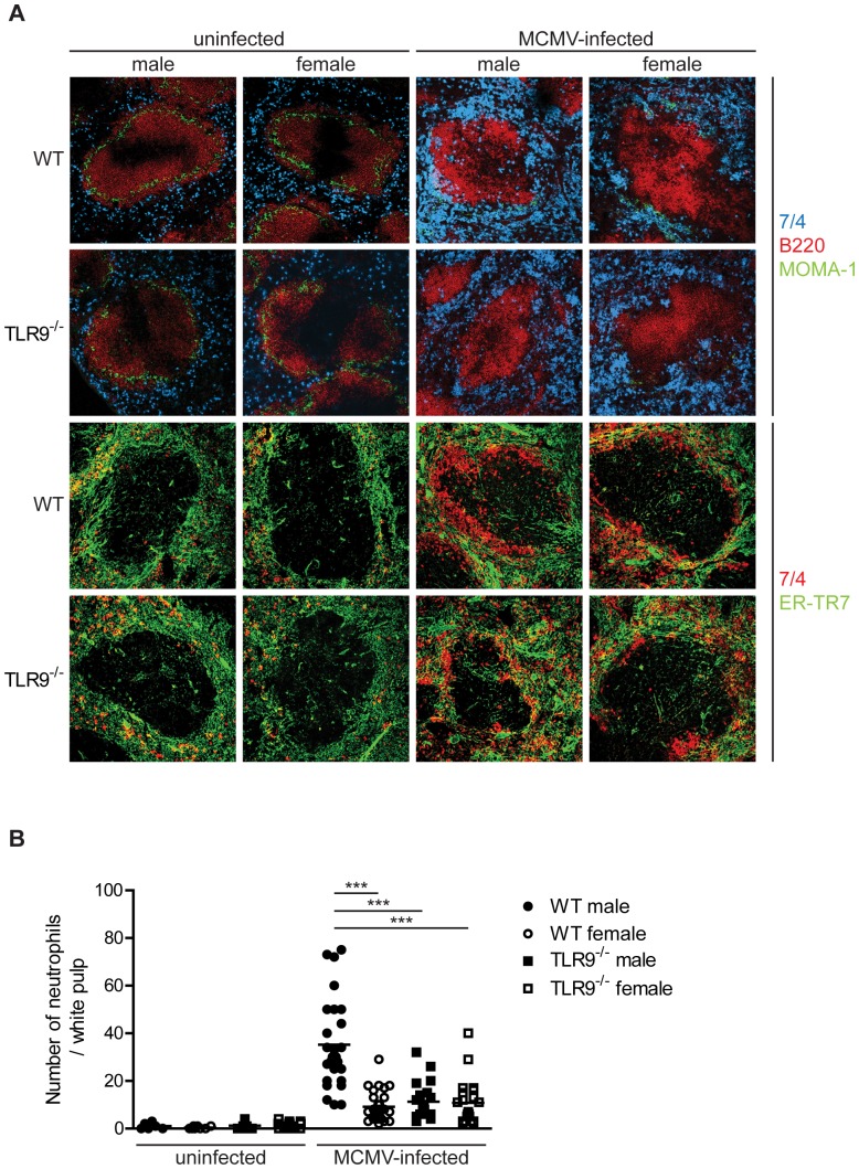
Figure 5. Increased number of neutrophils in the splenic white pulp of MCMV-infected WT male mice.
WT or TLR9−/− male and female mice were left untreated of infected with 1×10⁵ PFU of MCMV and 4 days later spleens were collected and processed for immunofluorescent analysis. (A) Murine spleen sections were incubated with antibodies specific to neutrophils (7/4, blue or red), B cells (B220, red), marginal metalophilic macrophages (MOMA-1, green) and reticular fibroblasts (ER-TR7, green). MCMV infection led to (A) a dramatic increase in the number of neutrophils that were present in the splenic red pulp and disappearance of the MZ metallophilic macrophages and (B) an increase in the number of neutrophils in the splenic white pulp areas compared to uninfected mice. These phenomena were more prominent in WT male than in WT female or TLR9−/− male and female mice. (B) Number of neutrophils per white pulp area were counted on slides stained in A. *** p<0.001. Data are representative of 9 mice per group.

