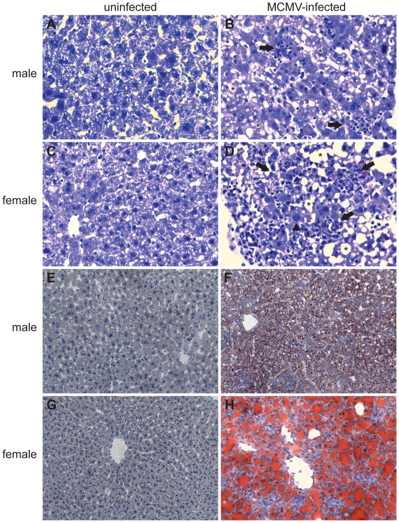
Figure 7. Increased liver inflammation and steatosis in MCMV-infected female mice.
(A) Livers were harvested and tissue sections were prepared from uninfected (A, C, E and G) or day 3 MCMV-infected male (B and F) and female (D and H) mice as described in Materials and Methods. Hematoxylin and eosin stained liver sections from uninfected (A and C) or MCMV infected (B and D) mice revealed inflammatory and necrotic foci in MCMV-infected mice. Female infected mice had increased inflammation and necrosis (more numerous and bigger foci) compared to male mice. Arrows identify inflammatory and necrotic foci, arrowhead shows an hepatocyte with an enlarged nucleous (karyomegaly) which contains large eosinophilic intranuclear inclusion and pheripheralized chromatin, asterisks indicate intracytoplasmic clear vacuoles. Accumulation of lipids (red stained lipid droplets) was detected by Oil red O staining (E–H) and was significantly more obvious in MCMV-infected female (H) versus male (F) liver sections, while in uninfected (E and G) mice was absent. Magnifications: A–D ×40 and E–H ×20. Data are representative of 2 independent experiments and with 3 mice per group.

