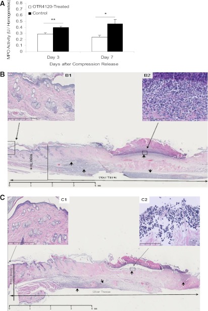
FIG. 3.
Effect of OTR4120 treatment on reducing neutrophil infiltration assessed by MPO activity assay (A) and hematoxylin and eosin (H&E)–stained histology (B and C). Representative H&E staining section on day 7 after compression release of a control ulcer (B) shows intense infiltration of neutrophils (arrows) (original magnification ×1.25; scale bar = 4 mm) in the ulcer area, and an OTR4120-treated ulcer (C) shows less intense infiltration of neutrophils (arrows) (original magnification ×1.25; scale bar = 4 mm). B and C, top left insets: Normal skin tissue, located 2 mm from the ulcer margin of a control ulcer (B1) and an OTR4120-treated ulcer (C1) (original magnification ×10; scale bar = 500 µm). B and C, top right insets: Neutrophil infiltration at higher magnification of a control ulcer (B2) and an OTR4120-treated ulcer (C2) (original magnification ×40; scale bar = 100 µm). Data are presented as means ± SEM. *P < 0.05 and **P < 0.01 indicate significant differences between the OTR4120-treated groups and control groups. (A high-quality color representation of this figure is available in the online issue.)

