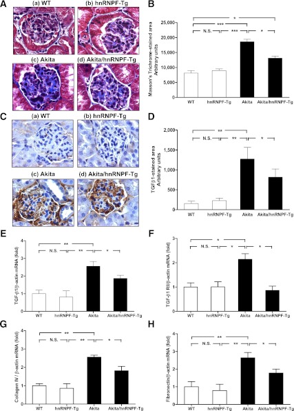
FIG. 5.
Overexpression of hnRNP F ameliorates glomerulotubular fibrosis and suppresses profibrotic gene expression in Akita Tg mice. A: Masson’s Trichrome staining of collagenous components expression in mouse kidneys: WT control mouse (a), hnRNP F-Tg mouse (b), Akita mouse (c), and Akita hnRNP F-Tg mouse (d). Original magnification ×600. B: Semiquantitative analysis of Masson’s Trichrome staining in glomerulotubular areas of kidney sections from WT control, hnRNP F-Tg, Akita, and Akita hnRNP F-Tg mice at week 20. C: Immunohistochemistry of TGF-β1 protein expression in mouse kidneys: WT control mouse (a), hnRNP F-Tg mouse (b), Akita mouse (c), and Akita hnRNP F-Tg mouse (d). Original magnification ×600. D: Semiquantitative analysis of TGF-β1 staining in kidney sections from WT control mice, hnRNP F-Tg mice, Akita mice, and Akita hnRNP F-Tg mice at week 20. RT-qPCR of TGF-β1 (E), TGF-β1 RII (F), collagen type IV 1α (G), and fibronectin (H) mRNA expression in RPTs of WT controls, hnRNP F-Tg, Akita, and Akita hnRNP F-Tg mice. Values are expressed as means ± SEM; N = 6. WT and hnRNP F-Tg mice, empty bars; Akita and Akita hnRNP F-Tg mice, filled bars. *P < 0.05; **P < 0.01; ***P < 0.005. (A high-quality digital representation of this figure is available in the online issue.)

