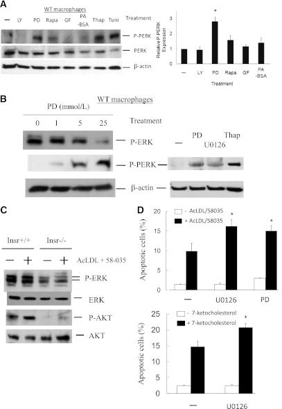
FIG. 2.
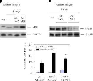
Inhibition of macrophage MEK signaling induces the UPR and promotes ER stress–associated apoptosis. A and B: Concanavalin A–elicited peritoneal WT macrophages were treated with inhibitors of PI3K (LY294002 [LY], 10 μmol/L), MEK (PD98059 or U0126, 10 μmol/L), mammalian target of rapamycin (rapamycin [Rapa], 2 μmol/L), protein kinase C (GF109203 [GF], 2 μmol/L), or palmitic acid–BSA (PA-BSA, 0.5 mmol/L) in DMEM with 10% FBS for 12 h. Protein lysate of macrophages treated with thapsigargin (Thap, 5 μmol/L) or tunicamycin (Tuni, 2.5 μg/mL) was used as positive controls. The levels of indicated proteins were determined by Western analysis. Data represent mean ± SEM. n = 3. *P < 0.05. C: Concanavalin A–elicited peritoneal macrophages isolated from Insr+/+ and Insr−/− mice were treated with or without AcLDL (100 μg/mL) + 58-035 (10 μg/mL) for 5 h. The expression of P-ERK, ERK, P-AKT, and AKT was determined by Western analysis. n = 3. D: WT peritoneal macrophages pretreated with or without the inhibitors of MEK (U0126 or PD98059) were incubated with or without AcLDL (100 μg/mL) and compound 58035 (10 μg/mL) or 7-ketocholesterol (40 μg/mL) at 37°C for ∼8 h. Apoptosis of macrophages was determined by annexin V staining. n = 3. *P < 0.05 for free cholesterol–loaded cells with vs. without the treatment of U0126 or PD98059 compounds. E and F: Concanavalin A–elicited peritoneal macrophages isolated from Insr+/+ (WT) and Insr−/− mice were infected with adenovirus either harboring lacZ (Ad-LacZ) or constitutively active MEK mutant (Ad-MEK) for ∼30 h. Macrophage protein extracts


