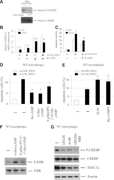
FIG. 4.
The activity of transcription factor CREBP downstream of MEK signaling is markedly reduced in insulin-resistant macrophages. A: Fresh peritoneal macrophages from either Insr+/+ or Insr−/− mice fed regular chow diet were cultured in DMEM with 10% FBS for 2 h. Cells were harvested and nuclei were fractionated. Nuclear P-CREBP and CREBP were determined by Western analysis with antibodies against the proteins as indicated. Fold change indicates the expression ratio of Insr−/− over Insr+/+. n = 3. *P < 0.05. B: WT or ob/ob macrophages were treated with AcLDL (100 μg/mL) and compound 58035 (10 μg/mL) with or without PKA activator Br-cAMP (10 μmol/L) for 8 h. Total RNA was then isolated for real-time QPCR analysis of SERCA2 mRNA expression. QPCR was performed in triplicate. n = 3. Averages of two independent experiments were shown. *P < 0.05 for WT vs. ob/ob cells with or without cholesterol loading. **P < 0.05 for ob/ob cells with vs. without Br-cAMP treatment. C: WT or ob/ob macrophages were treated as described in B. Apoptosis was determined by annexin V staining. n = 3. *P < 0.05 for free cholesterol–loaded WT vs. ob/ob cells. **P < 0.05 for free cholesterol–loaded ob/ob cells with vs. without Br-cAMP treatment. D: WT macrophages pretreated with or without Br-cAMP (200 μmol/L), 6-Bnz-cAMP (200 μmol/L), or 8-pMeOPT-2′-O-MecAMP (200 μmol/L) were incubated with AcLDL and compound 58035. Apoptosis was determined by annexin V staining. *P < 0.05 for free cholesterol–loaded cells with vs. without the treatment of cAMP analogs. n = 3. E: WT macrophages were incubated with AcLDL and compound 58035 with or without PKA inhibitors H-89 (5 μmol/L) or Rp-cAMPS (100 μmol/L). Apoptosis was determined by annexin V staining. *P < 0.05 for free cholesterol–loaded cells with vs. without the treatment of inhibitors. n = 3. F: WT macrophages were treated with or without 6-Bnz-cAMP (200 μmol/L) or 8-pMeOPT-2’-O-Me-cAMP (200 μmol/L) for 20 min. The levels of P-ERK and total ERK were determined by Western analysis. n = 3. G: WT macrophages were treated with or without U0126 (10 μmol/L) or H-89 (5 μmol/L) or in combination for 5 h. The levels of indicated proteins were determined by Western analysis. n = 3.

