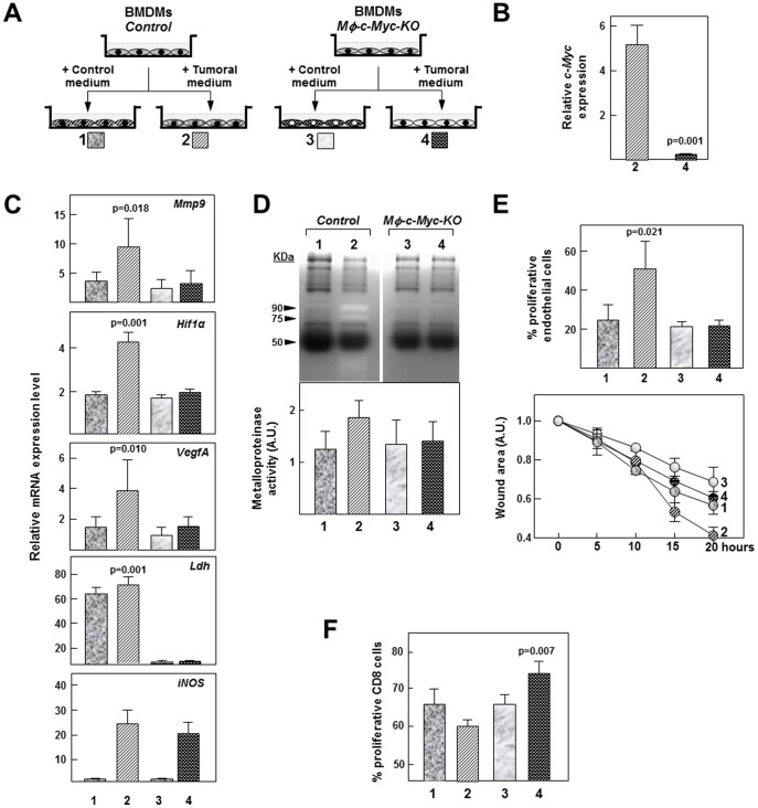
Figure 6. In vitro analysis of the pro-tumoral behavior of macrophages from control and Mφ-c-Myc-KO mice.
(A) Strategy for studying pro-tumoral behaviour of macrophages cultured in vitro in conditioned medium from control medium or tumor conditioned medium from B16 melanoma cells (n = 3 per genotype) (B) Expression levels of c-Myc mRNA in control and Mφ-c-Myc-KO BMDMs treated with tumor-cell–conditioned medium. (C) Transcript levels in BMDMs from both genotypes treated with control or tumor-cell–conditioned medium. (D) Top: Representative zymography analysis of metalloproteinase activity in supernatants from BMDMs treated with control or tumor-cell–conditioned medium. Bottom: Quantification of metalloproteinase activity determined by zymography analysis (n = 3 experiments). (E) Top: Quantification of endothelial cell proliferation 24 h after stimulation with supernatant from BMDMs treated with control or tumor-cell–conditioned medium. Bottom: Quantification of wound area 24 h after stimulation of scrape-damaged endothelial cultures with supernatant from BMDMs treated with control or tumor-cell–conditioned medium. (F) Quantification of CD8-T lymphocyte proliferation 24 h after stimulation with supernatant from BMDMs treated with control or tumor-cell–conditioned medium.

