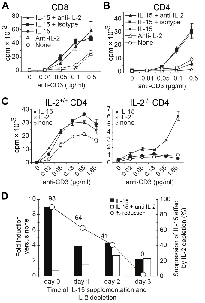
Figure 2. IL-15 growth factor activity depends on TCR-signals, but is IL-2-dependent only in CD4+ T cells.
(A) CD8+ or (B) CD4+ T cells were stimulated with anti-CD3 mAb and irradiated TdACs in the absence or presence of 3 ng/ml rhIL-15, or 10 µg/ml anti-IL-2 mAb (JES6-5H4) or isotype control, as indicated. On day 4, 3H-thymidine incorporation was measured. Results are from one of five experiments. (C) IL-2+/+ or IL-2−/− CD4+ cells were stimulated with anti-CD3 mAb and irradiated IL-2+/+ and IL-2−/− splenocytes, respectively, in the absence (white circles) or presence of 1 ng/ml IL-2 (crosses) or 3 ng/ml IL-15 (black circles). (D) CD4+ cells were stimulated for 5 days with 0.1 µg/ml anti-CD3 and irradiated TdACs. On day 0, 1, 2, or 3, IL-15 alone or IL-15 plus anti-IL-2 mAb was added. On day 4, proliferation was measured by 3H-thymidine incorporation and represented as bar graphs (with left Y-axis) as ratio of cpmIL-15/cpmnone (black bars) or cpmIL-15 plus anti-IL-2/cpmnone (white bars). The line represents the percentage of reduction of the IL-15 effect by IL-2 depletion as calculated using the following formula: [(cpmIL-15– cpmIL-15 plus anti-IL-2)/cpmIL-15]×100. Results are from one representative out of three experiments.

