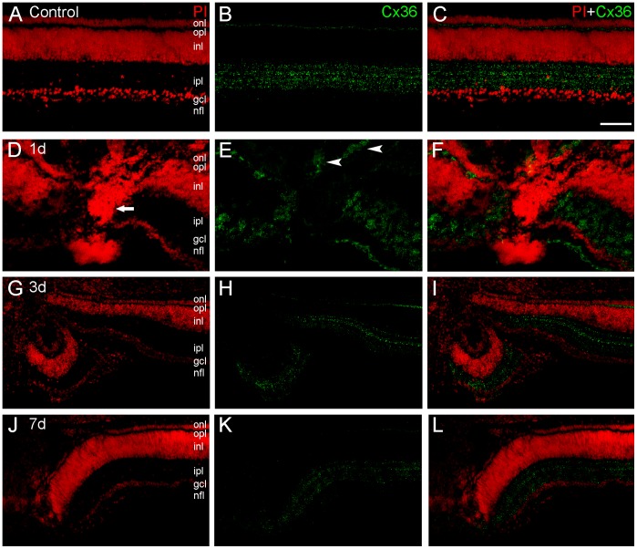
Figure 2. Connexin36 (Cx36) immunolabeling in transverse sections of the chick retina after mechanical lesions.
We determined spatial distribution of Cx36 protein (green) in retinas counterstained with propidium iodide (PI, red) in control retinas (A–C) and after 1-(D–F), 3-(G–I) and 7-days (J–L) of the mechanical lesion. Control retinas showed a pattern of Cx36 distribution similar to that described in previous studies, with punctate labeling in the inner plexiform layer (IPL) forming horizontal lines, and in the outer plexiform layer (OPL) as well. In the lesion focus it is possible to observe an increase of PI labeling evidencing apoptotic nuclei (arrows). In 1-day lesioned retinas, an unexpected labeling pattern was observed in the outer nuclear layer (arrowheads), revealing that cells in this layer accumulate Cx36 protein close to the lesion. In the 3- and 7-day lesioned retinas, in spite the course of the degeneration process, Cx36 punctate labeling remains visible, indicating the presence of this protein after in these time points after the lesion. Scale bar: 60 µm.

