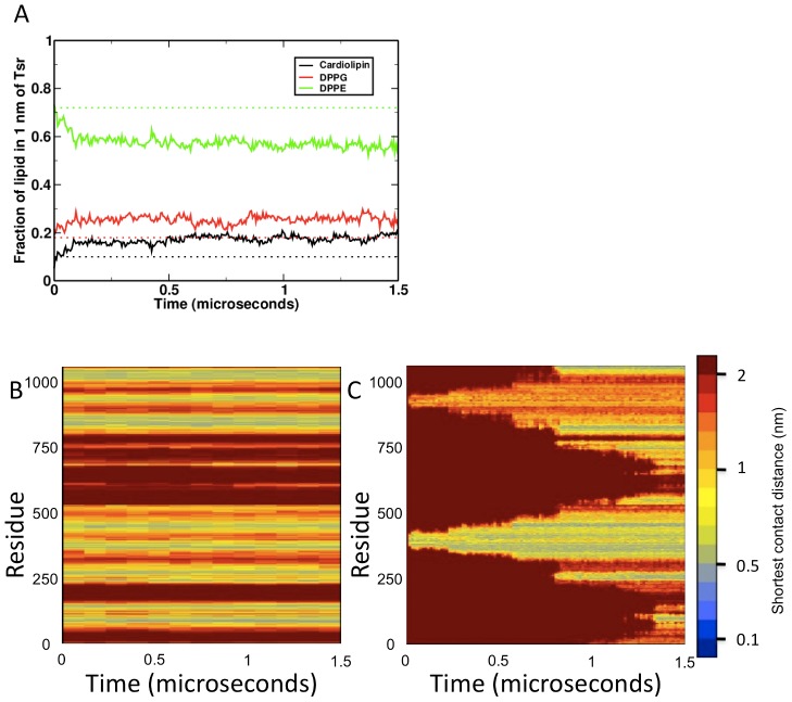
Figure 6. Interactions of proteins with their environment over time.
(A) Specific enrichment of anionic lipids (cardiolipin and DPPG) within a 1 nm distance of the proteins over time, relative to DPPE. (B) and (C) Example interaction fingerprints from trimer-of-dimers and dimer vesicle simulations. These show the shortest distance between a given residue in one dimer and any residue in the binding partner over time (based on the approach in [19]). (B) shows dimer-dimer interactions within a trimer-of-dimers, and (C) shows dimer-dimer interactions which appear over the course of the simulation. Over time, the interaction surface in (C) changed to mimic the stable, unchanging binding surface of the trimer-of-dimers model (B), indicating that the structure naturally oligomerises along this interface.

