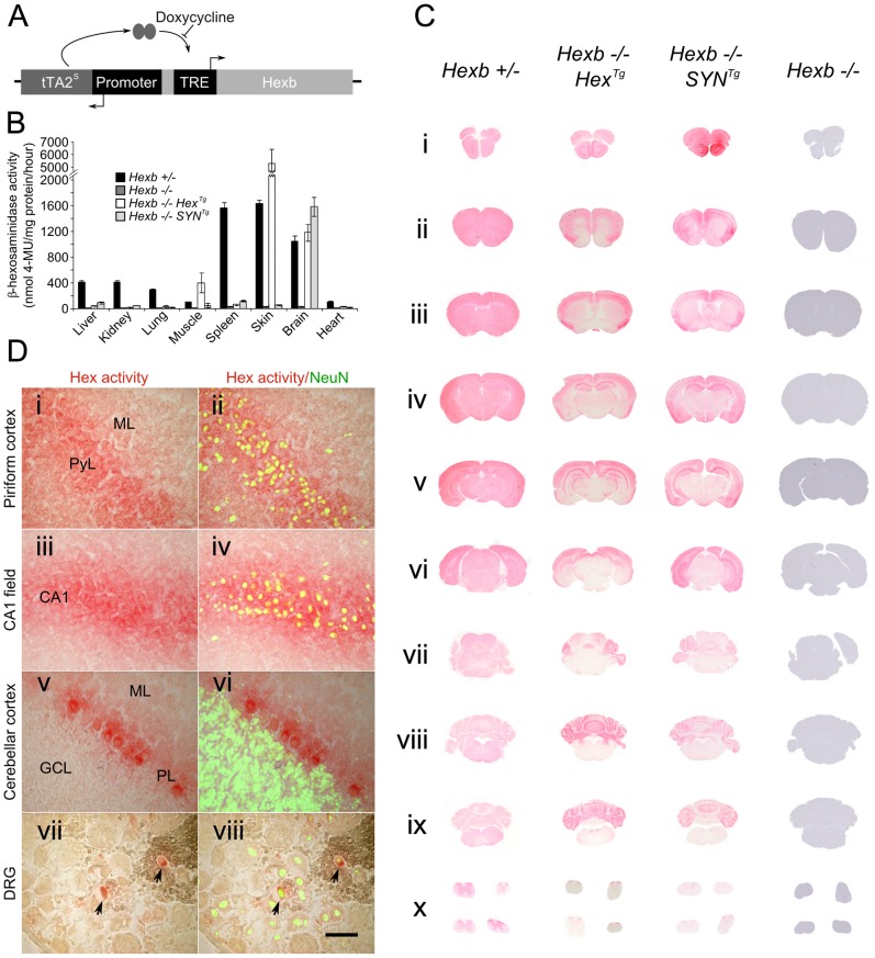
Figure 1. Expression of Hexb from two inducible constructs throughout the Sandhoff mouse neuraxis.
(A) Expression of transgenic Hexb from a single autoregulatory cassette was driven by either the mouse Hexb promoter (Hex line) or the human SYN1 promoter (SYN line). Tet-transactivator expressed from tTA2s coding sequence promoted expression of Hexb cDNA from tet-response elements (TRE). This was inhibited by doxycycline. (B) Expression of transgenic Hexb in different tissues was assessed with the MUG assay. β-hexosaminidase activity was found in the brain for both Hex and SYN lines, but also in the skin and skeletal muscle for the transgenic Hex mouse line. Bars = mean ± SEM. n = 3. Panel C shows expression of β-hexosaminidase activity in Hexb−/−HexTg and Hexb−/−SYNTg mice assessed by staining using the enzyme substrate naphthol AS-BI N-acetyl-β-glucosaminide (red staining). Strong expression of β-hexosaminidase activity is seen in the cortex (C i–vi) and the cerebellum (C vii–ix) of both lines. Weaker expression is seen in the diencephalon in the SYN line (C iv–v) while activity is very weak to absent in the mid (C vi–vii) and hindbrain (C vii–ix). (D) β-hexosaminidase activity staining was associated with neurons, shown by co-labelling with NeuN (green fluorescence) in the piriform cortex (i and ii), CA1 field of Ammon's horn (iii and iv), the cerebellar cortex (v and vi) and the dorsal root ganglia (vii and viii), where only some neurons expressed transgenic β-hexosaminidase activity. Images represent staining from Hexb−/−HexTg mice (DRG and cerebellar cortex) and Hexb−/−SYNTg mice (CA1 field and piriform cortex). ML = molecular layer, PyL = pyramidal layer, CA1 = CA1 field, PL = Purkinje neuron layer, GCL = granule cell layer. Scale bar = 50 µm.

