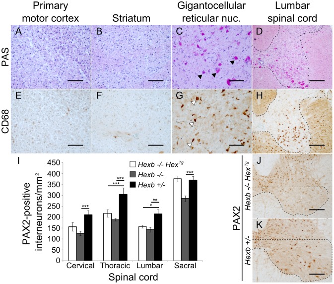
Figure 4. Localized glycolipid storage and microgliosis in Hexb−/−HexTg mice at humane endpoint.
PAS stained brain sections show regions of the cerebrum such as the primary motor cortex (A) and the striatum (B) are devoid of glycolipid storage that stains magenta. However, storage is a prominent feature in the hindbrain of the same animals. C and D show glycolipid storage in neurons of the brainstem (gigantocellular reticular nucleus) and in the spinal cord grey matter respectively (C, arrowheads; D, dashed line). (E–H) Staining for activated microglia is revealed by brown DAB staining for CD68 and coincides with storage (G, arrowheads show CD68 staining microglia; H, dashed line shows spinal grey matter). (I) PAX2-positive ventral horn interneurons were quantified for Hexb−/−HexTg, Hexb−/− (both humane endpoint) and Hexb+/− (one year old) animals (n = 6, 8 and 6. Bars = mean ± SEM. *, P<0.05; **, P<0.01; ***, P<0.001 – Bonferroni post hoc test). Both Hexb−/−HexTg and Hexb−/− animals showed loss of PAX2-positive neuron density in multiple regions of the ventral spinal cord compared with Hexb+/− animals. J and K show PAX2 stained lumbar spinal cord used for quantification. The dashed line encompasses the region quantified. Scale bars: A–C and E–G = 50 µm; D, H, J and K = 100 µm.

