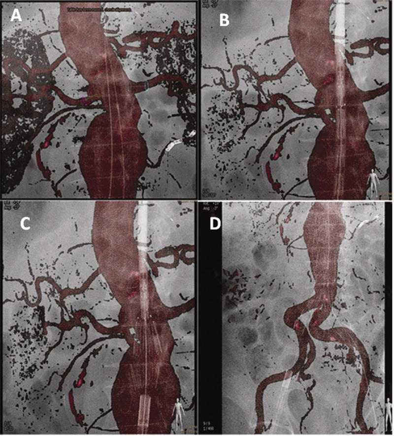
Figure 5.
Stent graft deployment for infra-renal abdominal aortic aneurysm using the pre-operative CTA as a 3D roadmap overlaid on the live fluoroscopy. Figure 5a Markers displayed on the renal arteries to be preserved (red and blue circles). One inferior right accessory renal artery was covered and one lower left accessory renal artery was embolized prior to stent graft deployment to avoid type II endoleaks. Two guidewires are visible in the projection of the aortic lumen. Figure 5b delineates positioning of the stent graft using the CTA as a roadmap enables a view of the aortic neck without requiring a standard angiogram. Figure 5c illustrates deployment of the first two struts of the stent graft under the 3D CTA roadmap control. Figure 5d shows the deployment of the entirety of the aorto-bi-iliac component of the stent graft. Note the deformation of the iliac arteries due to stiff guidewires and the stiffness of the stent-graft.

