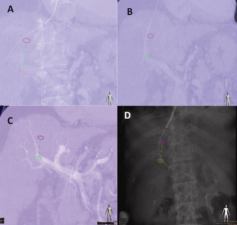
Figure 6.
A transjugular intra-hepatic portosystemic shunt (TIPS) procedure using the portal vein phase of a CT to guide catheterization of the portal vein with XperGuide needle path (Philips Healthcare Systems, Best NL). Figure 6a demonstrates guidewire positioned in the right hepatic vein and projected over of the needle path (green line flanked by green and pink circles). Figure 6b shows the needle positoned at the level of the right portal vein (note the small position shift due to the liver motion during breathing). An angiographic control after crossing with the 10 French long introducer is seen in figure 6c. Figure 6d delineates imaging following deployment of the covered stent between the right hepatic vein and the right portal vein.. Note the alignment of the stent relative to the planned needle path.

