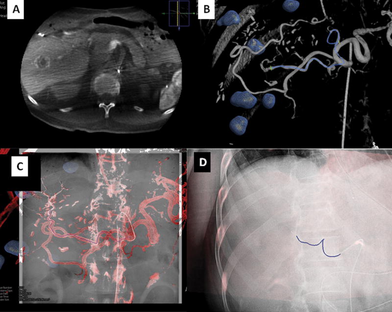
Figure 7.
Vascular application of CBCT: Figure 7a demonstrated a dual phase CBCT of the liver in a patient with metastatic colorectal carcinoma. The tumors are segmented in the same fashion as with the ablation platform (blue spheres). Tumor vessel supply may be segmented manually or automatically (figure 7b–c). The 3D roadmap can be superimposed on real-time fluoroscopy to navigate to the target vessel (blue line flanked by green crosses). It is possible to display the entire 3D roadmap or simply the vessel path (figure 7d). This 3D roadmap adjusts with table motion, change of C-arm position, and image magnification.

