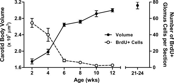
Fig 2.
Postnatal growth of the carotid body in Sprague-Dawley rats in terms of carotid body volume (filled symbols, n=8–13 per age) and the number of glomus cells that underwent mitosis in the preceding 24 hours (i.e., number of BrdU-positive glomus cells) (open symbols, n=3–5 per age). Although this experiment focused on growth between 2 and 12 weeks of age, carotid body volumes for 24–26 week old rats from another experiment (i.e., Control rats from Fig. 3B) are plotted for comparison. No effect of sex was detected, so data for males and females have been pooled. All values are mean ±SEM.

