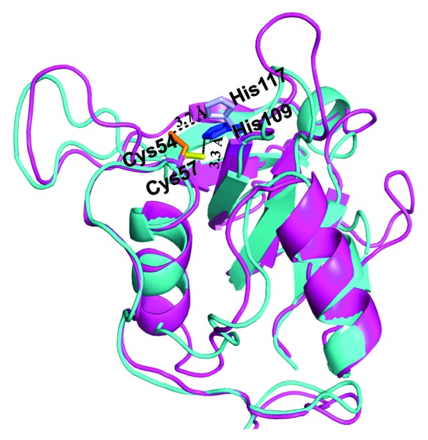
Figure 4. Structural comparison by superposition of the experimentally resolved 2K3A protein (cyan) and predicted CHAPK structure (magenta). Highly conserved active site residues are identified and labeled for both proteins. CHAPK Cys54 (orange) and His117 (light blue), 2K3A Cys57 (yellow), His 109 (blue). Figure created in PyMOL and analyzed in LSQMAN42 returning a value of 1.9 Å over 143 C-α atoms.
