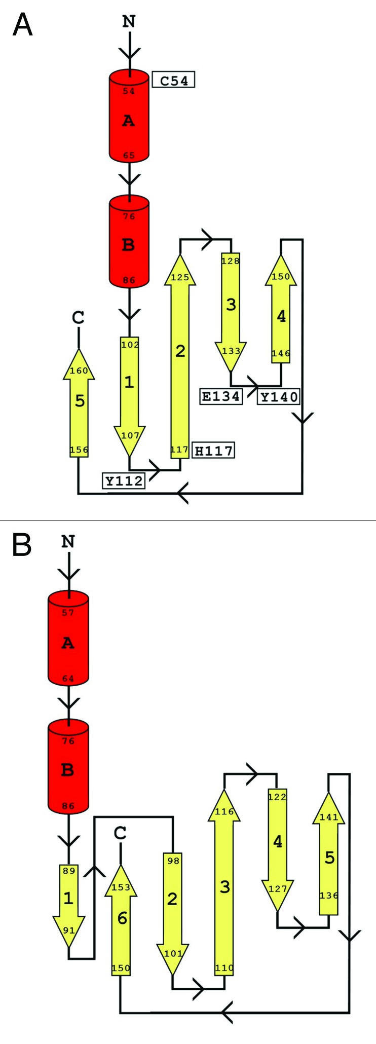
Figure 5. Topology maps for CHAPK and 2K3A proteins. Secondary structure was determined using DSSP. The topology maps show α-helices in red and the strands of the β-sheets in yellow. (A) shows the topology map for CHAPK while (B) shows the topology map for the Staphylococcus saprophyticus CHAP domain (PDB identifier 2K3A). Key residues in CHAPK are indicated in diagram (B).
