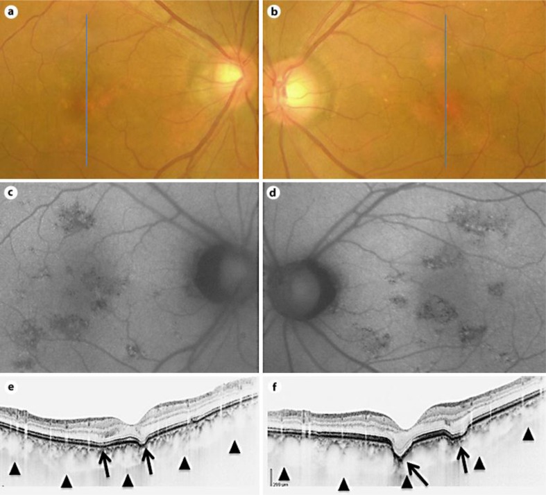
Fig. 1.
Findings in a 58-year-old man with bilateral focal choroidal excavation. a, c, e Images of the right eye. b, d, f Images of the left eye. a, b Fundus photographs on the initial presentation show RPE alterations beneath and adjacent to the fovea. c, d FAF image shows a focal area of hypoautofluorescence. e, f SD-OCT image with enhanced depth imaging through the fovea shows two concave-shaped conforming focal choroidal excavation (arrows) in both eyes. The excavations involved the outer retinal layers up to the ELM. The ELM band and the photoreceptor inner segment/outer segment junction can be seen at the excavation.

