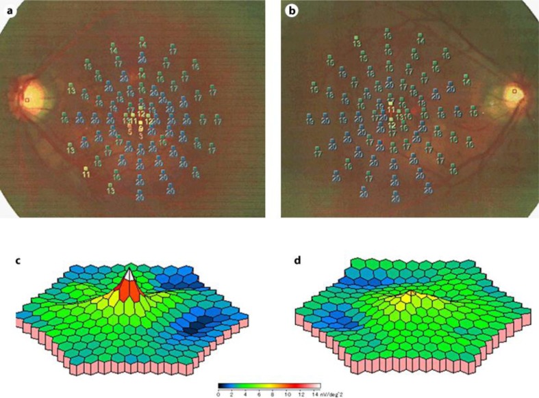
Fig. 2.
Case 1. a, c Images of the right eye. b, d Images of the left eye. a, b Microperimetry using MP-1 (Nidek, Japan) shows a decrease of retinal sensitivity in the foveal region in both eyes. c, d Results of mfERG using an LE-4100 (Tomey, Japan). Three-dimensional topographic map of the mfERG responses showing a decrease in the P1 response amplitude in the foveal region of the left eye.

