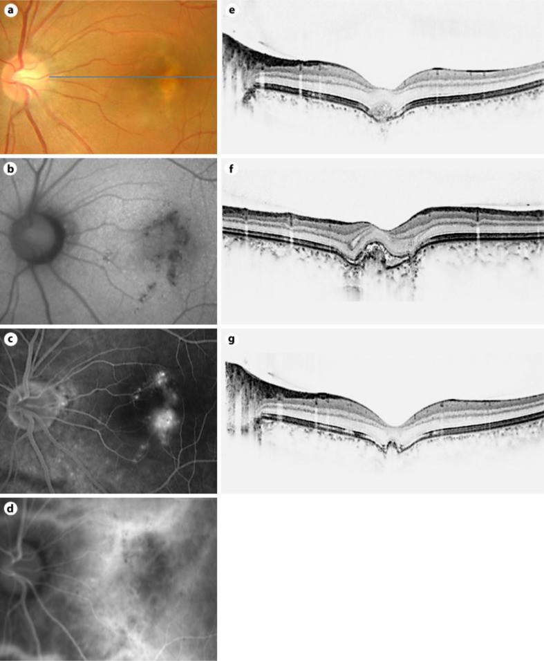
Fig. 3.
Findings in a 34-year-old woman with a focal choroidal excavation accompanied with type 2 CNV in the left eye. a Color photograph shows RPE alterations superior to the fovea. b FAF image shows an irregular area of hypofluorescence. c Fluorescein angiography in the late phase shows a classic CNV in the fovea. d Indocyanine green angiography in the late phase shows a classic CNV in the fovea. e SD-OCT at the initial examination shows a focal choroidal excavation with a type 2 CNV. The ELM band and the photoreceptor inner segment/outer segment junction are indistinct at the excavation. f SD-OCT scan taken 1 month after the initial presentation shows a type 2 CNV with subretinal fluid. g SD-OCT scan taken 5 months later shows a regression of the type 2 CNV after treatment with intravitreal bevacizumab injection.

