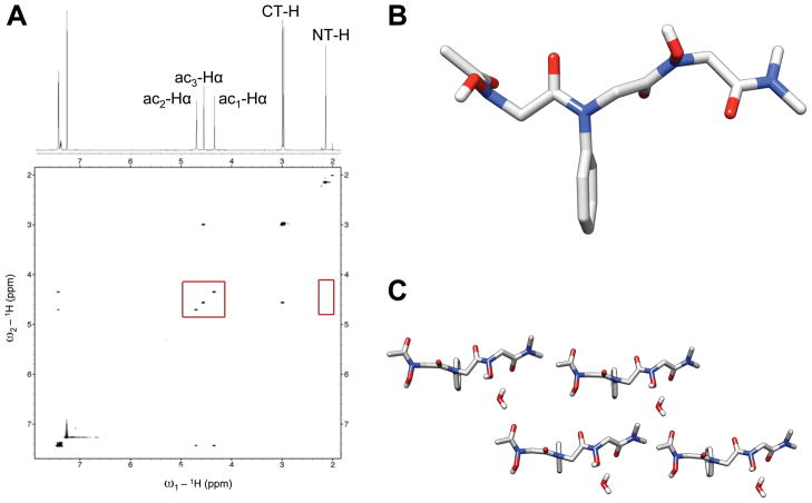
Figure 9.
(A) Partial 1H NMR and NOESY spectra of peptoid 4 in (600 MHz, CDCl3, 5 mM, 24 °C). The red boxes highlight the lack of an NOE between the NT-H and ac1-Hα backbone protons (right box), as well as the lack of an NOE between any of the ac-Hα backbone protons (left box). (B) View of the X-ray crystal structure of 4 without the co-crystallized water molecule. (C) A packing diagram of four molecules of 4 from the X-ray crystal data including water molecules. All hydrogens are omitted for clarity except for water molecules and amide side chain N-OHs.

