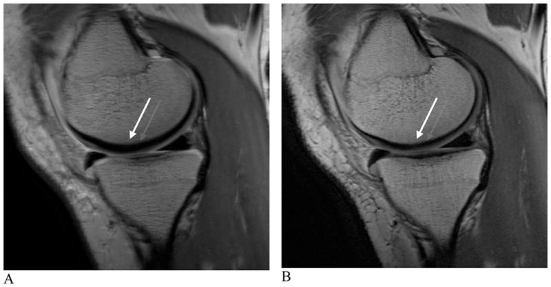
Figure 2. Proton density-weighted images of the healthy knee at 3.0 T.

A and B, sagittal images with bandwidths of 15 kHz (A) and 42 kHz (B). An approximate three-fold increase in bandwidth can significantly reduce chemical shift. The anatomy is more easily visualized and much sharper after the bandwidth increase (arrows, A, B).
