
Figure 3. T1-weighted fast spin-echo images of the healthy knee at 3.0 T.
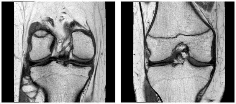
A and B, coronal images with TR/TE = 800/20 ms and echo train length of 4 resulted in an image with 33% of the average radiofrequency power limit compared with multi-slice spin echo. Slight blurring can be visualized due to the use of a short TE and multiple echoes.