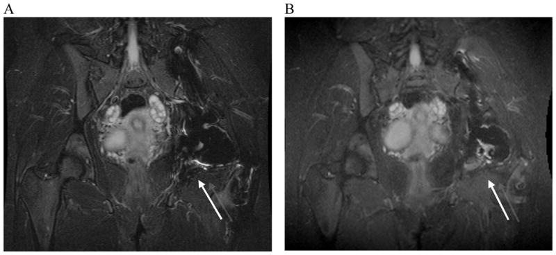
Figure 6.

Standard fast spin-echo inversion-recovery and SEMAC inversion recovery images of a patient with a history of chondrosarcoma and total hip replacement. A) 2D-FSE inversion recovery image. B) SEMAC inversion recovery image. The 2D-FSE inversion recovery image (A) shows artifact inferior to total hip replacement (arrow). The SEMAC inversion recovery image (B) shows an area of high signal in the bone marrow (arrow). High signal was suspicious of recurrent tumor.
